Design by{" "} Seo To Webdesign
Retinal Detachment
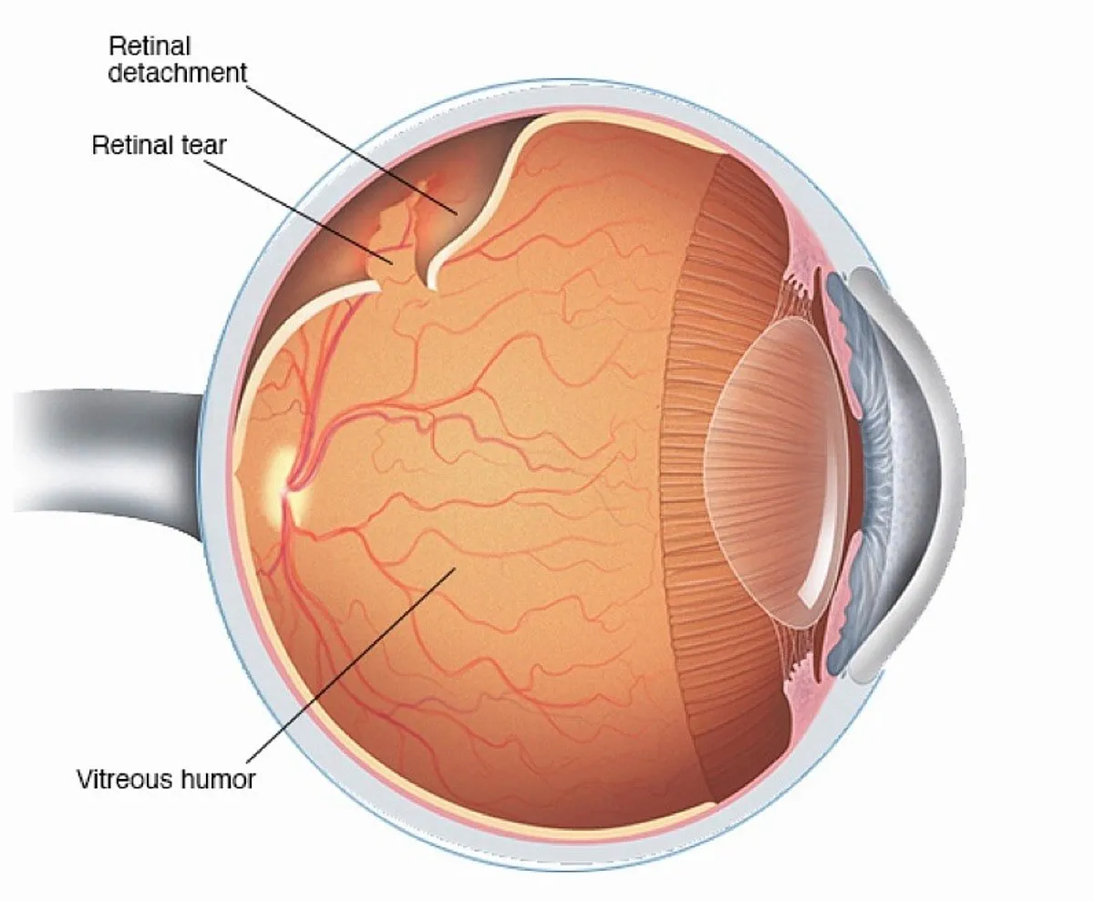
Retinal Detachment: Causes, Types, and Treatment
Retinal detachment is a critical eye condition that occurs when the retina—a light-sensitive tissue at the back of the eye—becomes separated from its supportive layer. This separation disrupts blood supply and retinal function, potentially leading to permanent vision loss if untreated. Prompt medical attention and diagnosis are essential for preventing serious outcomes.
Causes and Types of Retinal Detachment
Causes of Retinal Detachment
Retinal detachment can result from several factors, such as:
- Aging: Degeneration of the vitreous gel inside the eye can lead to retinal tears or holes.
- Eye Injuries or Surgeries: Trauma or surgical procedures may increase the risk of detachment.
- Medical Conditions: Diabetes and other diseases can damage retinal tissue, leading to detachment.
- Genetics: Family history of retinal detachment can predispose individuals to the condition.
Types of Retinal Detachment
Understanding the type of detachment is crucial for determining the appropriate treatment.
- Rhegmatogenous Retinal Detachment:
- The most common form caused by a tear or hole in the retina, allowing fluid to seep behind it. Aging, trauma, or retinal thinning are frequent contributors.
- Tractional Retinal Detachment:
- Scar tissue growth on the retina exerts traction, causing it to pull away from the underlying layers. This type is often associated with conditions like diabetic retinopathy.
- Exudative or Serous Retinal Detachment:
- Fluid accumulation beneath the retina occurs without tears or holes. It is often linked to inflammation, tumors, or macular degeneration.
Treatment Options for Retinal Detachment
Timely intervention is essential to prevent vision loss. Depending on the severity and type, treatments may include:
- Laser Photocoagulation: Uses lasers to seal retinal tears, preventing fluid from entering behind the retina.
- Cryotherapy: A freezing technique that promotes scarring to close tears and secure the retina.
- Pneumatic Retinopexy: Injects a gas bubble into the eye to push the retina back into place, followed by laser or cryotherapy.
- Scleral Buckling: Places a silicone band around the eye to relieve pressure and support reattachment.
- Vitrectomy: Removes vitreous gel and replaces it with gas or fluid to reattach the retina, ideal for complex cases.
Features
- Comprehensive Diagnostic Services: Advanced tools like OCT and ultrasound for accurate diagnosis.
- Modern Surgical Techniques: Access to the latest procedures like scleral buckling and vitrectomy.
- Expert Ophthalmologists: Experienced specialists ensuring personalized care.
- 24/7 Support: Round-the-clock assistance for consultations and emergencies.
FAQ
-
1. What are the early signs of retinal detachment?
Symptoms include sudden flashes of light, an increase in floaters, or a shadow/curtain effect over your vision.
-
2. Is surgery always required for retinal detachment?
Not always. Early-stage tears or holes may be managed with laser or cryotherapy. Advanced cases typically require surgery.
-
3. How long is the recovery time after surgery?
Recovery can vary, but most patients regain stability within a few weeks to months, depending on the procedure.
-
4. Can retinal detachment be prevented?
Regular eye exams, managing health conditions like diabetes, and protecting your eyes from injury can reduce risk.